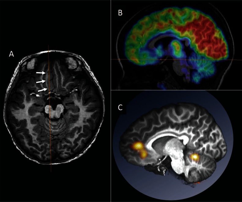
Fig. 3.
Multimodal presentation in the anatomical space. A 6-year-old girl with the right medial frontal lobe epilepsy is presented. Abnormally-thickened cortex was noted in the right medial frontal lobe (arrows in A). FDG-PET in the sagittal section revealed the area of glucose hypometabolism corresponding to the lesion (B). Subtraction ictal SPECT co-registered to MRI (SISCOM) images were registered on the patient’s MRI and presented in the same section with the FDG-PET (C). Ictal hyperperfusion was found to occur in the dorsal part of the lesion. Surgery was planned to remove both the lesion and the area of ictal hyperperfusion. Spatial co-registration of multimodal images is useful in surgical planning.

