
Figure 1.
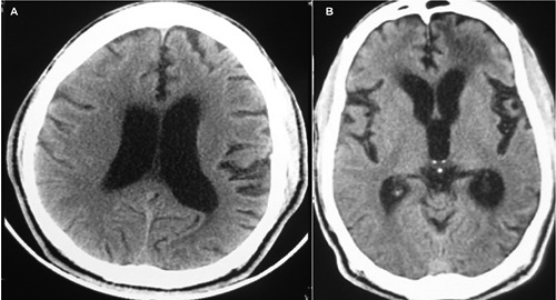
A) Axial non contrast computed tomography (CT) brain done five months prior to presentation showing cortical atrophy and hydrocephalus; B) axial non contrast CT brain on admission again showing hydrocephalus, with bilateral temporal atrophy, with hypodensities in the subcortical frontal region of both hemispheres, but more prominent on the left than right.