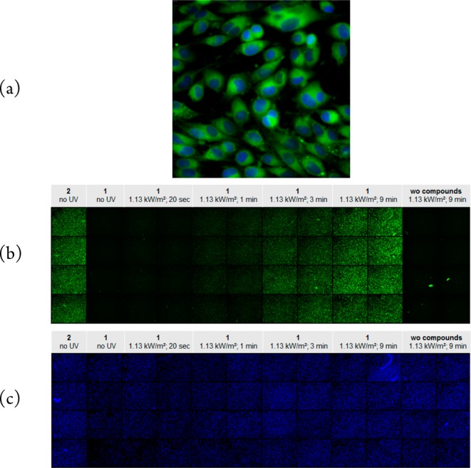
Figure 5.

(a) SKMEL28 cells were incubated with dabrafenib_photo (2). The inhibitor was applied at 100 μM concentration and is shown in green. The cell nuclei were counterstained with 1 μg/mL DAPI and are marked in blue. The microscopic image was taken with 60× magnification. (b) SKMEL28 cells were seeded in 48 wells of a 96-well plate. The cells in the first column were incubated with dabrafenib_photo (2) without UV exposure. The cells in the columns 2 to 10 were incubated with dabrafenib (1) and exposed to increasing dosage of UV light at 365 nm (see captions above the columns). The cells in the last two right columns were not incubated with any compound but just irradiated with the highest UV amount. The overview image of the plate consists of single well images taken with 10× magnification. (c) The same cell plate from (b). All cell nuclei in the plate were counterstained with 1 μg/mL DAPI and are marked in blue.
