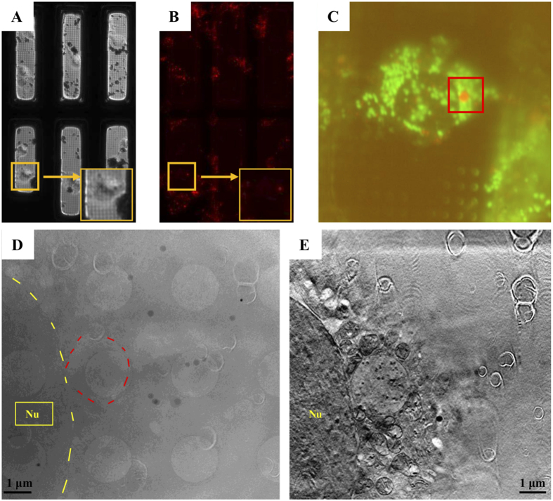
Figure 1. Sample preparation for cryo-soft X-ray tomography (SXT).
RBL-2H3 cells were grown on gold grids coated with Quantifoil holey-carbon films. The cells were prelabeled with fluorescent dye and frozen with a plunge freezer in liquid ethane. (A,B) Cryo-brightfield and cryo-fluorescence images of cells were captured with a 10x objective lens on the cryo-fluorescence screening system at −150 °C for screening of the samples. (C) Fluorescent images of the cryo-cells were captured with a 100x objective lens in the vacuum chamber to search for granular structures at U41-SGM beamline TXM endstation, BESSY II. Mitochondria (green) and granules (red) are visible. (D) An image captured by SXT was coordinated with the fluorescent dye stained granule (circle labeled) shown in (C). (E) A reconstructed SXT image by IMOD to show morphological structures of the organelles. Nu: nucleus.

