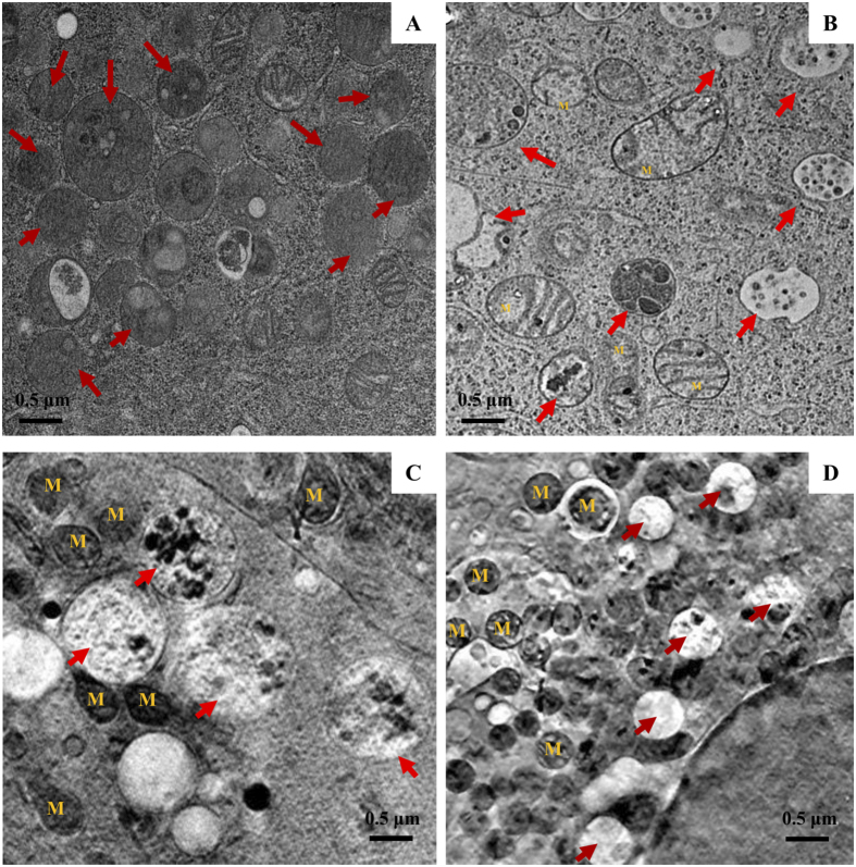
Figure 2. Granular morphology during mast cell degranulation.
RBL-2H3 cells were sensitized with anti-DNP IgE and then stimulated with or without DNP-BSA for 30 min. (A,B) Transmission electron microscopy (TEM) images of cells either not stimulated (A) or stimulated with DNP-BSA (B) followed by ultrathin section from Spurrs’ resin embedded cells. (C,D) Reconstructed images of SXT of cells non-stimulated (C) or stimulated with DNP-BSA (D). Arrows indicate granules and M indicates mitochondrion.

