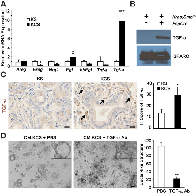
Figure 4.

Deletion of SMO in fibroblasts promotes ADM via TGF-α. (A) Quantitative real-time PCR analysis of known EGFR-activating ligands. n = 3. Error bars represent means ± SD. (B) Western blot of TGF-α in CM; SPARC was used for a loading control. (C) IHC and quantitation for TGF-α; the graph represents the quantification of the H score for each genotype. Bar, 25 μm. n = 3. Error bars represent means ± SD. (D) Images and quantitation of ductal-like structures in 3D ADM formation assay with pretreatment of KCS CM with TGF-α-neutralizing antibody. The insets show a higher magnification view of representative cellular structures. The graph represents the quantitation of ductal-like structures. Bar, 40 μm. n = 3. Error bars represent means ± SD. (*) P < 0.05; (**) P < 0.01; (***) P < 0.001.
