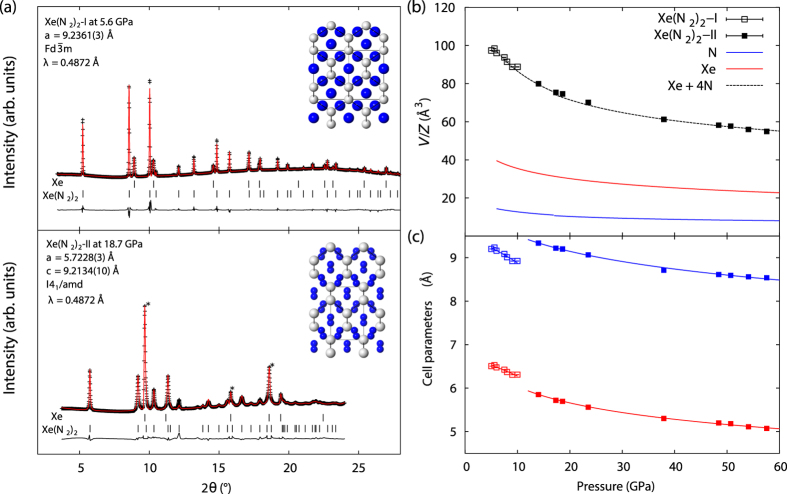
Figure 1.
(a) Powder X-ray diffraction patterns at 5.6 and 18.7 GPa used for Rietveld refinement. Below 14 GPa, Xe(N2)2 adopts a face-centered cubic structure, space group  , a = 9.2361(3) Å denoted Xe(N2)2-I. At 14 GPa and above Xe(N2)2 undergoes a transition to a body-centered tetragonal structure, I41/amd, with unit-cell dimensions a = 5.7228(3), c = 9.2134(10) Å at 18.7 GPa. Peaks corresponding to Xe (marked with *) were excluded from the profile used in the refinement. Insets show crystal-structure projections for both phases, phase I is rotated to view down the face diagonal 〈110〉 highlighting structural similarity to phase II. Freely rotating N2 molecules in phase I are represented by blue spheres, whilst in phase II blue spheres represent atoms in aligned N2 molecules; (b) Equation-of-state data for Xe(N2)2 compounds. Pressure-volume per Z data for Xe phases is indicated by red lines32, N2 phases by blue lines6. Black squares indicate volume per Z data for Xe(N2)2 phases I and II, dashed black line indicates the calculated volume for stoichiometry Xe + 4N from atomic volume data; (c) Response of unit-cell dimensions to applied pressure for Xe(N2)2, phase I data are shown for unit-cell length a (blue open squares) and d〈110〉 (red open squares), phase II data is plotted for unit-cell lengths a (red closed squares) and c (blue closed squares). Solid lines indicate fitted linear Birch-Murnaghan linear equations of state?

