Figure 2.
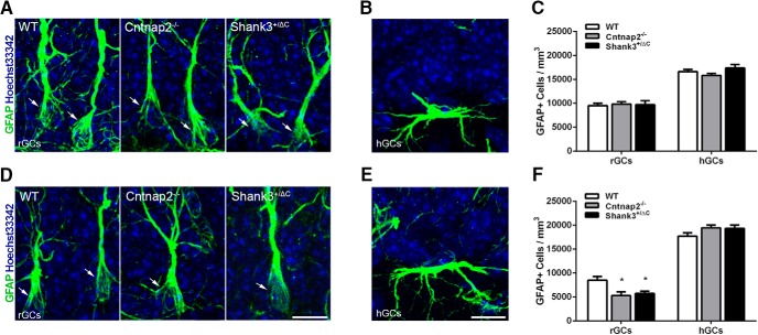
Cntnap2−/− and Shank3+/ΔC mice have reduced numbers of GFAP-labeled radial glial progenitor cells in the ventral dentate gyrus of the hippocampus A, D, Representative images of radial glial cells (rGCs) immunolabeled with astrocyte marker GFAP (green) and counterstained with Hoechst 33342 (blue) from the dorsal (A) and ventral (D) SGZ. Arrows point to GFAP+ radial glial cells. Scale bar, 10 µm, applies to all frames. B, E, Representative images of horizontal glial cells (hGCs) from the dorsal (B) and ventral (E) SGZ. Scale bar, 10 µm, applies to all frames. C, F, Quantification of the density of GFAP-labeled cells with radial glial morphology and horizontal morphology in the dorsal (C) and ventral (F) SGZ. Error bars represent the SEM. *p < 0.05.

