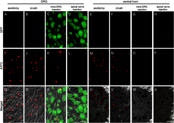
Figure 7. Expression of activating transcription factor 3 (ATF3) and green fluorescent protein (GFP) in the dorsal root ganglia (DRG) and spinal cord after spinal nerve injection (injection group) in comparison with sciatic nerve transection (axotomy group), spinal nerve crush (crush group), and intra-DRG injection (intra-DRG injection group).
Double-labeling immunofluorescence staining was performed for GFP (A–H) and ATF3 (I–P) in the DRG (1st, 2nd, 3rd and 4th columns) and spinal cord (5th, 6th, 7th, and 8th columns) in the axotomy (1st and 5th columns), crush (2nd and 6th columns), intra-DRG injection (3rd and 7th columns) and spinal nerve injection (4th and 8th columns) groups. Fluorescence staining and dark field images were merged (U–X). GFP was strongly expressed in the ipsilateral DRG (C,D,S,T) but was absent in the ventral horn (G,H,W,X) of the injection group. There were no GFP(+) neurons in the axotomy and the crush group (A,B,E,F,Q,R,U,V). ATF3 expression was evident in the ipsilateral DRG and ventral horn of the axotomy, crush and intra-DRG injection groups (I–K,M,N,Q–S,U,V), in contrast to the spinal nerve injection group (L,T,P,X). Bar, 50 μm.

