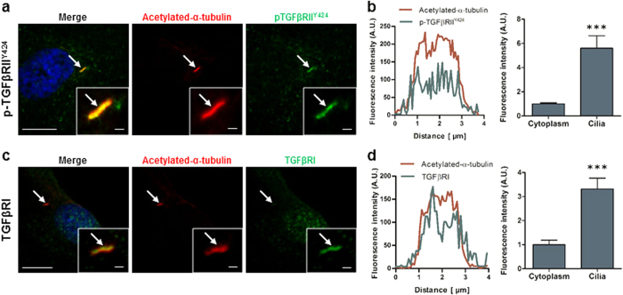
Figure 2. TGFβ receptors localise to the primary cilia in human MSC.
(a,b) Immunocytochemistry analysis of the cilia (identified by arrows) using anti-acetylated-α-tubulin in red, constitutively active TGFβRII (p-TGFβRIIY424) (a) and TGFβRI (b) in green and nucleus in blue (DAPI). Scale bar 5 μm and 1 μm in the enlarged pictures. (c,d) Fluorescence intensity analysis of the levels of p-TGFβRIIY424 (c) and TGFβRI (d). Intensity profile of Acetylated-α-tubulin (red) and receptors (green) along the cilia (left panel) and analysis of the average intensity in the cilia and cytoplasm (right panel). Statistical analysis using Student’s t-test, ***p < 0.001.

