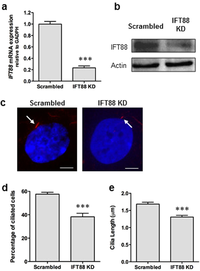
Figure 3. Knockdown of IFT88 in hMSCs impairs cilia formation/maintenance.

(a,b) Efficiency of IFT88 siRNA transfection on IFT88 mRNA expression analysed using qPCR (a) and Western blot (b). Expanded Western blot can be seen in Supplemental Figure S3. (c) Immunocytochemistry analysis of acetylated-α-tubulin (red) and DAPI (blue) showing shortened primary cilia (identified by arrows) after IFT88 KD. Scale bar 5 μm. (d,e) Analysis of the percentage of ciliated cells (d) and cilia length (e) after IFT88 KD. Statistical analysis using Student’s t-test. ***p < 0.001.
