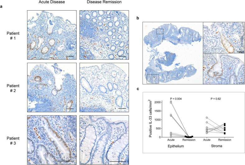
Figure 3. IL-33 is present in epithelial crypts in acute ulcerative colitis.
Panel (a) shows colonic biopsies from 3 representative patients with acute UC treated with infliximab (anti-TNF) to disease remission. Endoscopic biopsies were taken from the most inflamed colonic regions pre- infliximab therapy (acute disease), and repeated from the same region at disease remission. Biopsies were formalin fixed, paraffin embedded with immunoenzymatic staining for IL-33 (brown) using monoclonal mouse antibody (Nessy-1). IL-33 positive cells are seen in epithelial crypts during acute disease. At disease remission no epithelial staining for IL-33 was seen. Cell nuclei (blue) counterstained with hematoxylin. Scale bar = 0.1 mm. Panel (b) shows a representative whole biopsy from acute ulcerative colitis (x40 magnification) stained as above, whilst boxes show high-power magnification. Scale bar = 0.05 mm. Panel (c) shows quantification of IL-33 positive cells per mm2 in individual patients (n = 9) in the epithelium and lamina propria.

