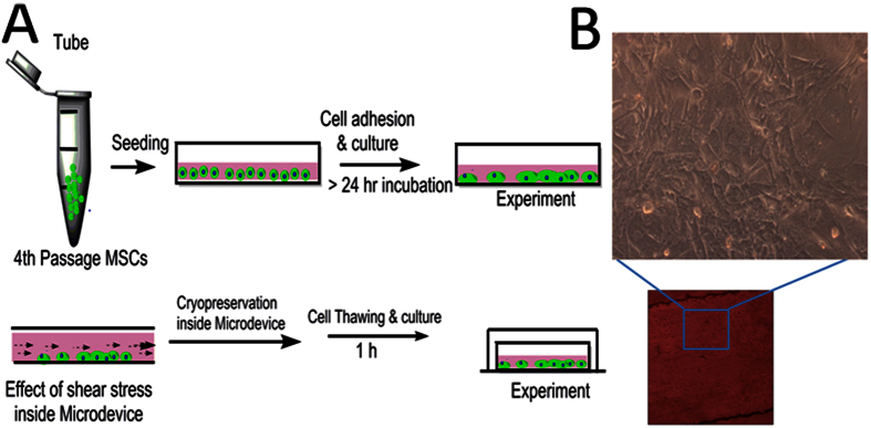
Figure 10.
Illustration of cryopreservation strategy used for MSCs inside microfluidic chamber (A) Schematic representation of cell seceding and subsequent cryopreservation protocol (B) Phase contrast image of MSCs inside microfluidic chamber and it enlarge image with 10X magnification obtained from confocal microscope.

