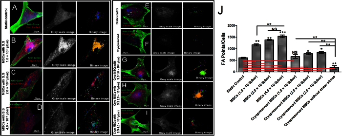
Figure 3. Vinculin associated focal point adhesions: Distribution of actin filaments and vinculin in human mesenchymal stem cells, showing cytoskeletal organization.
Actin filament, vinculin and nucleus are pseudocolor in green, red, and blue respectively. (A) 8th day, MSCs seeded in microchannel without any shear stress. (B) 8th day MSCs seeded in microchannel under 1 × 10⁻³ μbar. (C) 8th day MSCs seeded in microchannel under 2 × 10⁻³ μbar. (D) 8th day MSCs seeded in microchannel under 4 × 10⁻³ μbar. (E) Static Control (F) MSCs seeded in microfluidic device with cryopreservation treatment. Cryopreserved MSCs after 8 days of (G) 1 × 10⁻³ μbar, (H) 2 × 10⁻³ μbar, (I) 4 × 10⁻³ μbar shear stress inside microchannel. (J) Quantitative difference in focal points in MSCs at various shear stresses compare to static control. (NS: > 0.05, ** < 0.01, *** < 0.005).

