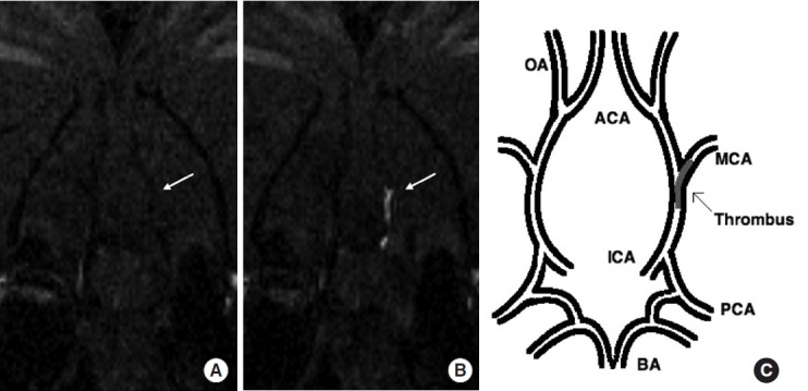
Figure 1.

Coronal slice from 3D T1-weighted images at the level of the middle cerebral artery (MCA) origin. The EP-2104R-enhanced image (B) clearly identifies the thrombus (arrow) that was not visible on the image acquired before EP-2104R injection (A). Pictorial representation of the location of the thrombus within the cerebral arterial tree (C). BA indicates basilar artery; PCA, posterior cerebral artery; ICA, internal carotid artery; OA, olfactory artery; ACA, anterior cerebral artery. Figure and caption reprinted from Uppal et al. Stroke (2016) [21] with permission.
