
Fig. 1.
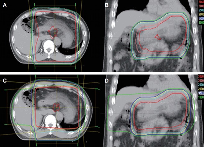
Axial (A) and coronal (B) computed tomography (CT) images of two-dimensional plan for radiotherapy with anterior-posterior opposing two fields and axial (C) and coronal (D) CT images of three-dimensional plan for radiotherapy with four coplanar fields.