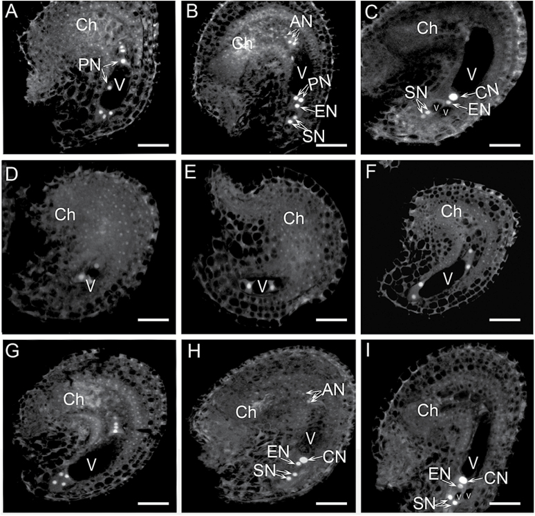
Fig. 1.
FG development in stage 12 flowers of wild-type and rid1-2/+ mutant plants. (A–C) Three stages of FG development. The embryo sacs at the FG 5 (A), FG6 (B), and FG7 (C) stages in pistils of wild-type stage 12 flowers. (D–I) Six stages of FG development. The embryo sacs at the FG2 (D), FG3 (E), FG4 (F), FG5 (G), FG6 (H), and FG7 (I) stages in pistils of rid1-2/+ stage 12 flowers. The developmental stages of FG were determined according to Christensen et al. (1997), and the nuclear position was examined by detecting their autofluorescence (for details see the Methods section). AN, antipodal nucleus; Ch, chalazal end; CN, central cell nucleus; EN, egg cell nucleus; PN, polar nucleus; SN, synergid nucleus; V, vacuole. Scale bars = 10 μm.

