Fig. 4.
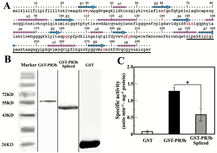
Specific chitinase activity of native and alternatively spliced PR3b. (A) Conserved α-helixes (blue arrows), β-sheets (pink bars), and amino acids (red characters) of PR3b compared with the catalytic cores of GH18 family chitinases (Hurtado-Guerrero and van Aalten, 2007). The region affected by alternative splicing is underlined with a black line. (B) Purified proteins of GST-tagged PR3b and GST separated by SDS-PAGE gel. ‘GST-PR3b’ is the GST-tagged protein of native PR3b; ‘GST-PR3b Spliced’ is the GST-tagged protein of spliced PR3b. Marker is the protein standard ruler. The picture shown is a combination of representative lanes from gels with serial elutions of the purified proteins. (C) Enzyme-specific activity of GST-tagged native and alternatively spliced PR3b. One unit equals 1 μmol of released 4-MU. GST is used for the control reaction. Values are the average of three replicates. Error bar, mean ±SD. The asterisk indicates a significant difference between the paired data sets (P <0.05, Student’s t test).

