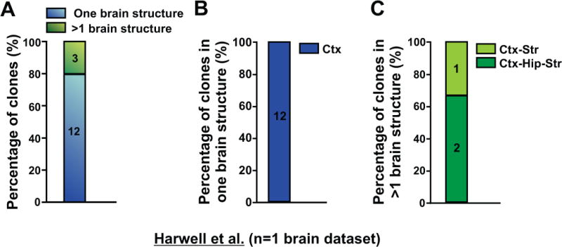
Figure 3. MGE/PoA-derived interneuron clones in Harwell et al. are mostly restricted to the cortex; See also Table S2.

(A) Quantification of the percentage of clones that are either restricted to one brain structure (blue) or span more than one brain structure (green). (B) Quantification of the percentage of clones restricted to one brain structure (n=2 hemispheres). (C) Quantification of the percentage of clones spanning more than one brain structure (n=2 hemispheres). Ctx, cortex; Hip, hippocampus; Str, striatum.
