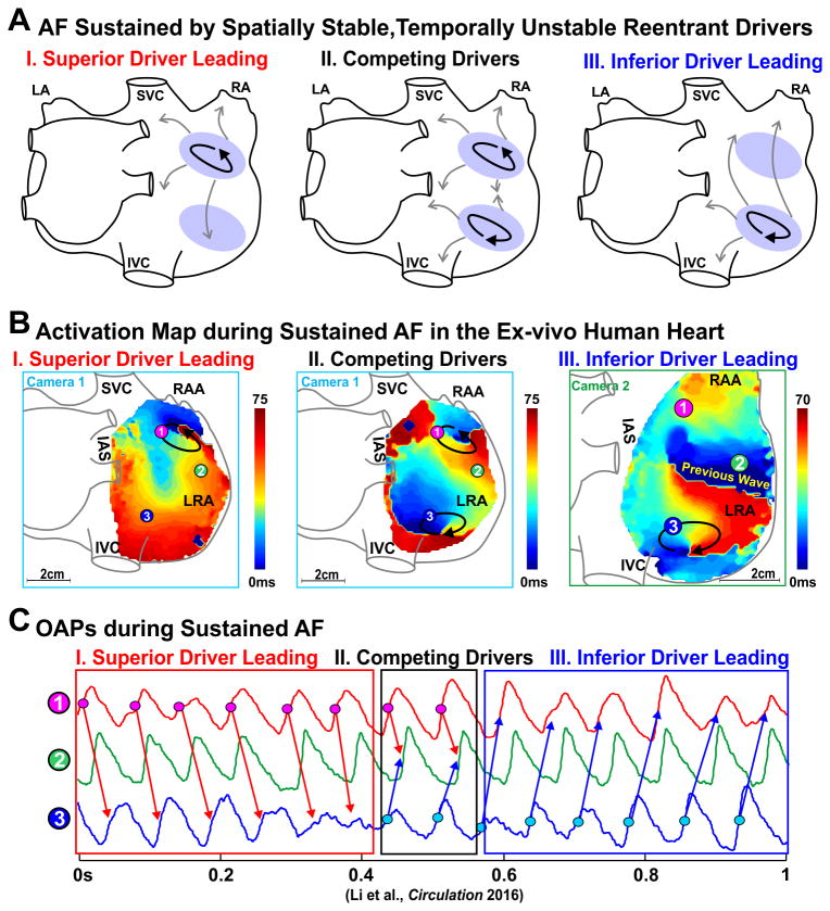
Figure 4.
AF maintained by competing drivers during sustained AF in the ex-vivo human heart. (A) Schematic of two spatially stable AF drivers competing in the RA. Pink oval, black arrows, and grey arrows represent the AF driver region, path of reentry, and fibrillatory conduction, respectively. (B) Activation maps showing AF driven first by a superior and then an inferior spatially stable AF driver. Arrows indicate the location and direction of AF reentrant drivers. (C) Optical action potentials (OAPs) from superior driver, inferior driver, and the middle right atrial free wall. Abbreviations as in Figure 3. From Li et al.3 with permission.

