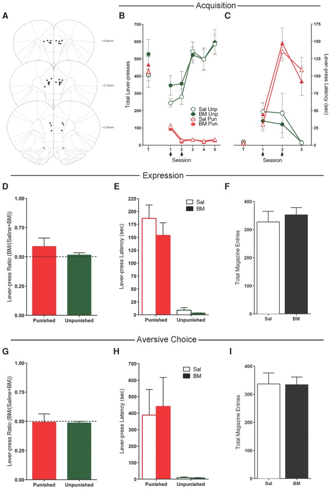
Figure 3.
Effects of prelimbic cortex (PL) inactivations. (A) PL cannula placements as verified by Nissl-stained sections. Black dots represent the ventral point of the cannula tract, indicated on coronal sections adapted from Paxinos and Watson (2007). (B) Mean ± SEM lever-presses on the punished and unpunished levers during the last day of lever-press training (T) and punishment acquisition (sessions 1–5). Arrows indicate days that rats received infusions of either saline (n = 7) or baclofen and muscimol (BM) (n = 8) immediately prior to the session. (C) Mean ± SEM latency to initially press the punished and unpunished levers (averaged across trials) during punishment acquisition. (D) Mean ± SEM lever-press ratios of BM on lever pressing during punishment expression (n = 15). (E) Mean ± SEM latency to initially press the punished and unpunished lever (averaged across trials) during punishment expression after infusions of saline or BM. (F) Mean ± SEM magazine entries during punishment expression after infusions of saline or BM. (G) Mean ± SEM lever-press ratios of BM on lever pressing during aversive choice (n = 15). (H) Mean ± SEM latency to initially press the punished and unpunished levers during choice test after infusions of saline or BM. (I) Mean ± SEM magazine entries during choice test after infusions of saline or BM.

