Figure 3.

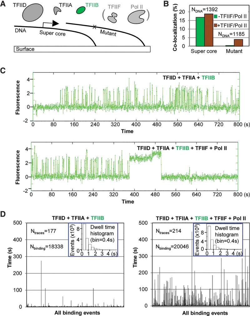
Change of TFIIB dynamics in the presence of TFIIF–Pol II. (A) Scheme: Fluorescently labeled TFIIB (4 nM; TMR) was mixed together with TFIID and TFIIA in the absence or presence of Pol II–TFIIF and incubated in the imaging chamber for binding to immobilized DNA templates. (B) Colocalization of TFIIB signals with the supercore or mutant promoter-containing DNA templates in response to the addition of TFIIF and Pol II. Results were obtained using the same set of DNA molecules incubated with two protein mixtures (first without TFIIF and Pol II and then with TFIIF and Pol II) for 13 min (800 sec). (C) A representative TFIIB fluorescence time trace from the same supercore DNA template in the absence (top) or presence (bottom) of TFIIF and Pol II. The transient-to-stable transition in the binding pattern occurred on approximately one-third of all DNA molecules bound by TFIIB in the presence of Pol II–TFIIF. (D) All TFIIB-binding events are represented by bars (height depicts residence time) to highlight the long events, which are vastly outnumbered by the short binding events in the histograms (insets).
