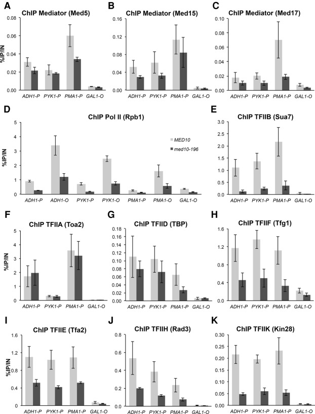
Figure 3.
ChIP analysis of PIC components in med10-196. Cells were grown to exponential phase at 30°C on YPD medium and then transferred for 90 min to 37°C. The immunoprecipitated protein is indicated together with the complex to which it belongs: Mediator tail (Med5) (A), Mediator tail (Med15) (B), Mediator head (Med17) (C), Pol II (Rpb1) (D), TFIIB (Sua7) (E), TFIIA (Toa2) (F), TFIID (TBP) (G), TFIIF (Tfg1) (H), TFIIE (Tfa2) (I), TFIIH core module (Rad3) (J), and TFIIH kinase module (Kin28) (K). All proteins, except Rpb1, were tagged with HA or TAP (see the Supplemental Material). Immunoprecipitated DNA was amplified with primers corresponding to ADH1, PMA1 and PYK1 ORF (O) or promoters (P). P1 primers listed in Supplemental Table S3 located close to upstream regulatory regions were used for Mediator ChIP experiments, and P2 primers located close to core promoters were used for Pol II and GTF ChIP experiments. GAL1 ORF was used as a negative control, since it is repressed in glucose-supplemented rich medium. The mean values and standard deviation (indicated by error bars) of three independent experiments are shown.

