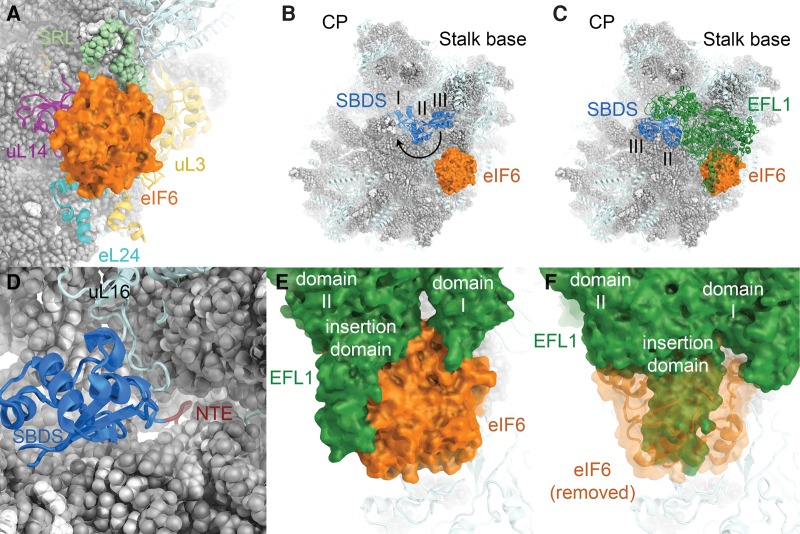
FIGURE 8.
Insights into the binding of eIF6 to the 60S subunit and its release by SBDS and EFL1. (A) Structure of eIF6 bound to the 60S subunit (PDB ID 4V8P). (B) Structure of the 60S-eIF6-SBDS complex (PDB ID 5AN9). SBDS domains are numbered. 60S subunit visualization based on the yeast 60S subunit (PDB ID 4V88). SBDS closed-to-open transition indicated by an arrow. (C) Visualization of the 60S-eIF6-SBDS-EFL1 complex (PDB ID 5ANB). (D) The SBDS N-terminal extension (NTE, dark red) enters the polypeptide tunnel near the PTC. For clarity, only the N-terminal domain I of SBDS is shown. (E) EFL1-eIF6 interactions in the 60S-eIF6-SBDS-EFL1 complex. EFL1 domains I and II as well as an insertion domain in domain II are indicated. (F) In the post-eIF6 release conformation, the position of EFL1 is incompatible with eIF6 binding due to extensive overlap (PDB ID 5ANC).

