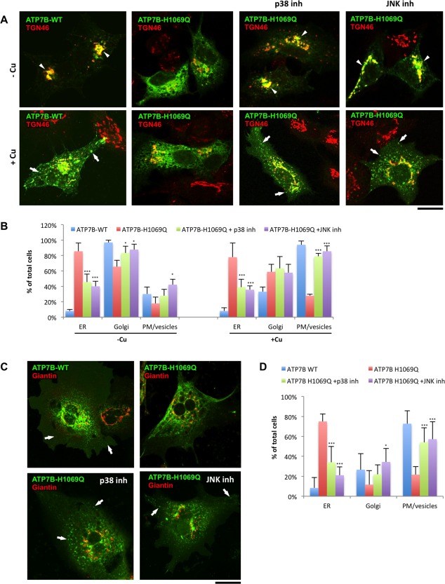
Figure 6.

Suppression of p38 and JNK corrects localization and trafficking of ATP7BH1069Q mutant in hepatic cells. (A) HepG2 cells were infected with Ad‐ATP7BWT‐GFP or Ad‐ATP7BH1069Q‐GFP, incubated overnight with 200 μM BCS, and fixed or incubated for an additional 2 hours with 100 μM CuSO4. In response to Cu, ATP7BWT (left column) traffics from the Golgi (arrowheads in upper panels) to the PM and vesicles (arrows in lower panels), while ATP7BH1069Q (second column) is retained in the ER under both low‐Cu and high‐Cu conditions. p38 or JNK inhibitors were added to the cells 24 hours before fixation (as indicated in the corresponding panels). Fixed cells were then labeled for TGN46 and visualized under a confocal microscope. Both p38 and JNK inhibitors corrected ATP7BH1069Q from the ER to the Golgi (arrowheads in upper panels) under low‐Cu conditions and to the PM and vesicles (arrows in lower panels) upon Cu exposure. (B) Cells were treated as in (A). The percentage of cells (average ± standard deviation, n = 10 fields) with ATP7B signal in the ER, Golgi, or PM/vesicles was calculated. Both p38 and JNK inhibitors reduced the percentage of cells exhibiting ATP7BH1069Q in the ER independently from the Cu levels and increased the number of cells in which ATP7B was corrected to the Golgi under low‐Cu conditions and to the PM and vesicles upon Cu stimulation. (C) Primary hepatocytes isolated from mouse liver cells were infected with Ad‐ATP7BWT‐GFP or Ad‐ATP7BH1069Q‐GFP and then incubated with 100 μM CuSO4 for 2 hours. p38 or JNK inhibitors were added to the cells 24 hours before fixation (as indicated in the corresponding panels). Fixed cells were then labeled for giantin and visualized under a confocal microscope. Both p38 and JNK inhibitors corrected ATP7BH1069Q from the ER to the PM and vesicles (arrows). (D) Primary hepatocytes were treated as in (C). The percentage of cells (average ± standard deviation, n = 10 fields) with ATP7B signal in the ER, Golgi, or PM/vesicles was calculated. Both p38 and JNK inhibitors reduced the percentage of cells exhibiting ATP7BH1069Q in the ER and increased the number of cells in which ATP7B was corrected to the PM and vesicles upon Cu stimulation. Scale bars = 5 μm (A) and 5.3 μm (C). Abbreviation: inh, inhibitor.
