
Figure 1.
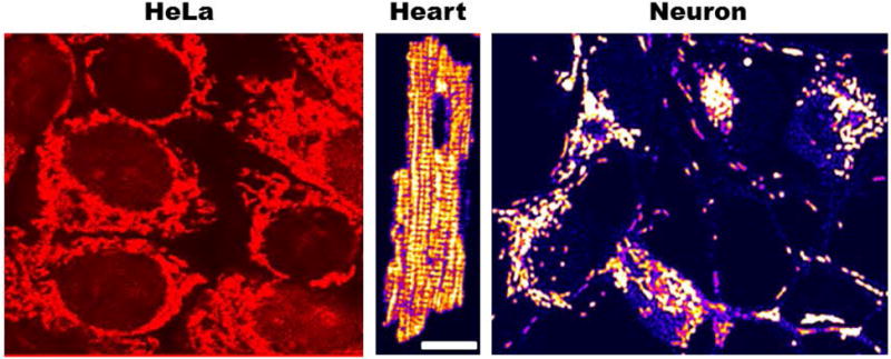
Morphological appearance of intracellular mitochondrial networks labeled with the cationic potentiometric dye tetramethylrhodamine ethyl ester (TMRE) used to monitor mitochondrial membrane potential. Notice the different architecture exhibited by mitochondrial networks in HeLa, cardiomyocyte and cortical neuron cells ranging from reticular (Hela and neuron) to lattice-like (ventricular cardiomyocyte).