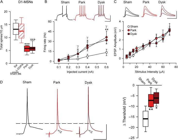
Figure 1.
Synaptic transmission and neural excitability are altered in PD and dyskinesia in direct striatal projection neurons. (A) Number of dendritic spines in 10 µm of D1-MSNs. (B) Number of action potential (AP) as a function of injected current intensity in D1-MSNs. Top, representative examples of firing rate at 0.4 nA of injected current. Scale bar = 50 mV/50ms. (C) Evoked-EPSP amplitude values across stimulus intensity in D1-MSNs. Top, representative examples of evoked EPSPs at 20, 40, and 60 µA. Scale bar 10 mV/20ms. (D) Illustrative traces of APs evoked by synaptic stimulation in each experimental condition in D1-MSNs (left) Summary of changes in the threshold of synaptically evoked APs; (*γP < 0.05, **γγP < 0.005 parkinsoninan or dyskinetic mice respectively vs. sham-lesioned; one-way ANOVA).

