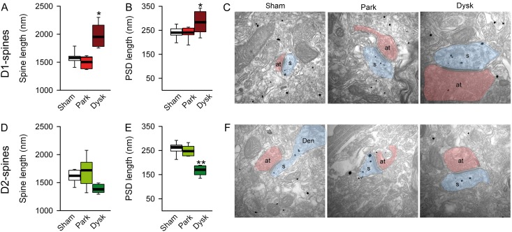
Figure 3.
PSD length is decreased in D2-MSNs in dyskinesia. EM analysis of spine length (A) and PSD length (B) in D1-MSNs. (C) Representative images of spines from D1-MSNs in each experimental condition. Spine length (D) and PSD length (E) of D2-MSNs. (F) Representative EM illustration of spine from D2-MSNs (*P < 0.05, **P < 0.005 vs. sham-lesioned; one-way ANOVA). at: axon terminal; s: spine.

