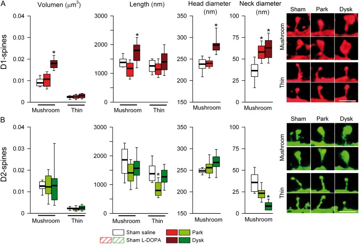
Figure 6.
Morphology of mushroom spines is modified selectively in D1-MSNs in dyskinetic mice. (A) Quantification of volume and length of mushroom and thin spines and head and neck diameter of mushroom spines of D1-MSNs. Right: Representative high magnification of mushroom (top) and thin (bottom) spines from D1-MSNs. Scale bar = 0.75 µm. (B) Analysis of the dendritic spines in D2-MSNs. Right: Representative mushroom (top) and thin (bottom) spines from D2-MSNs. Scale bar = 0.75 µm (*P < 0.05 vs. sham-lesioned; Kruskal–Wallis test).

