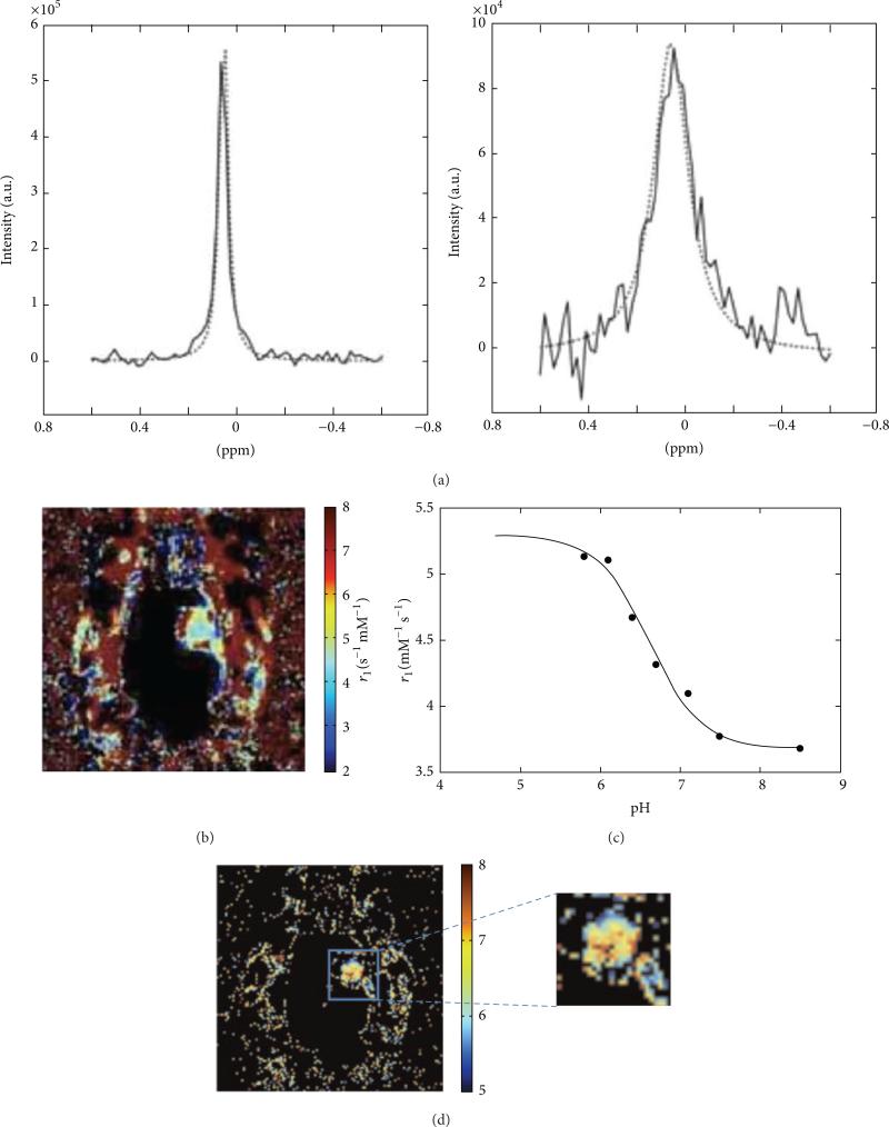
Figure 12.
Relaxation-based MRI of Gd-DOTA-4Amp and DyDOTP can measure tumor pHe. (a) The change in water linewidth before injection (left) and after injection (right) is used to estimate the concentration of the agent. (b) A parametric map of the r1 relaxivity of the agent in a glioma model is obtained from a T1-weighted MR images and the concentration of the agent. (c) The r1 relaxivity of the agent is pH-dependent, (d) which can be used to convert the r1 relaxivity map to a pH map (color scale bar shows pH units). Reproduced from [53] with permission.

