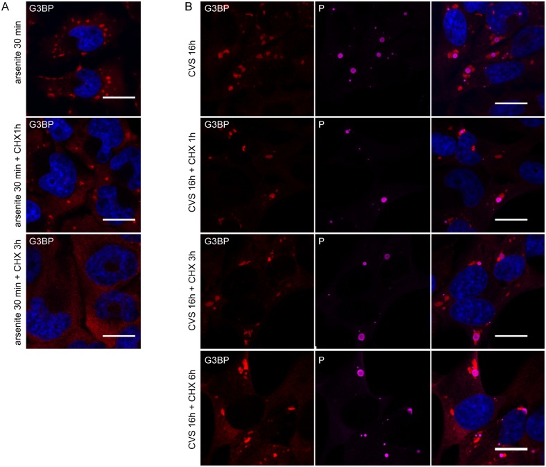
Fig 5. SGs induced by arsenite are disrupted by the protein synthesis inhibitor CHX whereas RABV-induced SGs are not.
(A) U373-MG cells were treated with arsenite (0.5mM) for 30 min and subsequently were either left untreated or treated with CHX for another 1 h or 3 h (as indicated) before processing for IF. Cells were then stained for G3BP1. DAPI (blue) was used to stain the nuclei (merge). The scale bars correspond to 15 μm. (B) U373-MG cells were infected with CVS for 16 h before IF (top panel) or were treated with CHX at 16 h p.i for 1 h or 3 h or 6 h as indicated and were then processed for IF to detect P and G3BP proteins. Staining with secondary antibodies and DAPI was carried as in Fig 1. The scale bars correspond to 15 μm.

