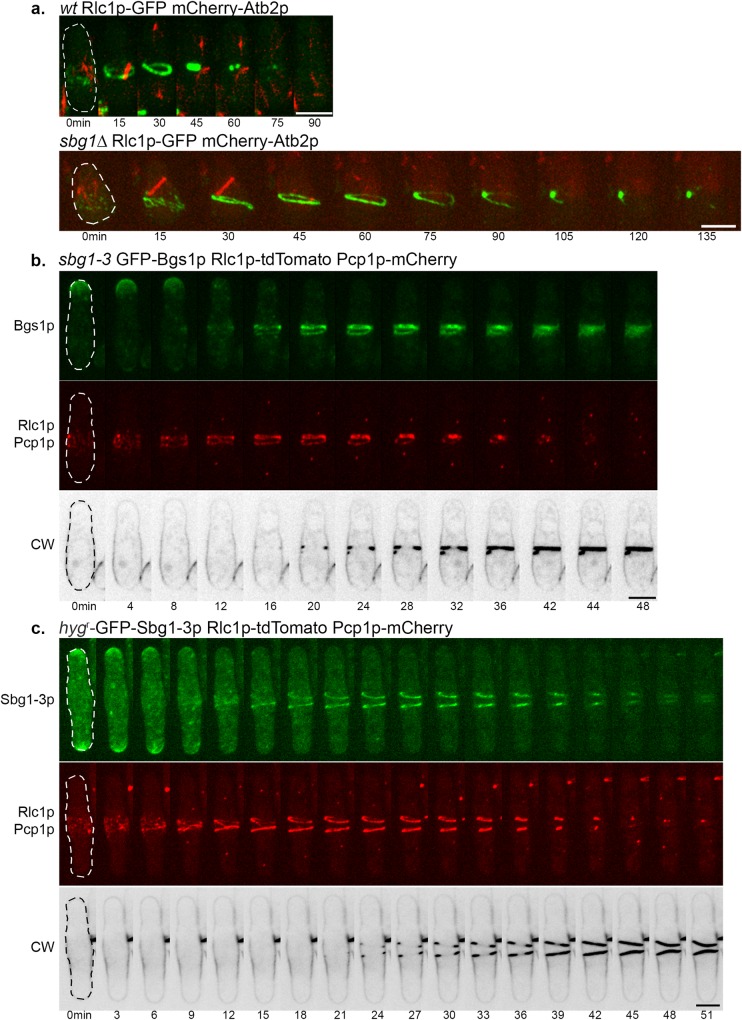
Fig 7. Actomyosin ring dynamics in sbg1 mutants.
(A) Maximum Z projection spinning disk confocal images of germinated cells of the indicated genotype from diploid sbg1Δ/sbg1+ mCherry-Atb2p Rlc1p-3GFP (MBY9156). Green, Rlc1p-GFP. Red, mCherry-Atb2p. (B) Maximum Z projection spinning disk confocal montage of the indicated strain (sbg1-3 GFP-Bgs1p Rlc1p-tdTomato Pcp1p-mCherry—MBY9390) after 6 hr at 36°C. Green, GFP-Bgs1p. Red, Rlc1p-tdTomato Pcp1p-mCherry. Calcofluor White (CW) images were acquired as single medial plane images and are inverted for fluorescence. 0min indicates time of spindle body duplication. Defects observed in 29.1%±7.9% cells (n = 3, at least 45 cells). (C) Maximum Z projection spinning disk confocal montage of the indicated strain (hygr-GFP-Sbg1-3p GFP-Bgs1p Rlc1p-tdTomato Pcp1p-mCherry—MBY9389) after 6 hr at 36°C. Green, hygr-GFP-Sbg1-3p. Red, Rlc1p-tdTomato Pcp1p-mCherry. Calcofluor White (CW) images were acquired as single medial plane images and are inverted for fluorescence. 0min indicates time of spindle body duplication. Defects observed in 37.4%±6.7% cells (n = 2, at least 20 cells). Scale bar 5μm.

