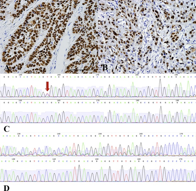
Fig 3. p53 IHC and TP53 exons 5–8 Sequencing.
(A, B) Accumulation of abnormal p53 protein in tumor cell nuclei. (C, D) An atlas of direct sequencing of TP53 exons 5–8. Upper photographs show different classifications of mutants, and lower photographs are normal controls; TP53 exons 5–8 point mutation are marked with red arrow in picture C; Heterozygous TP53 exons 5–8 frame-shift mutations are shown in picture D.

