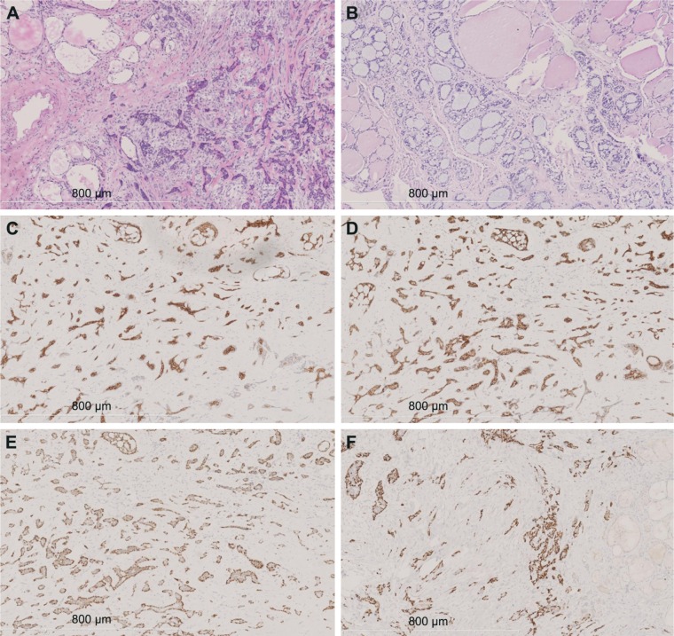
Figure 2.
Immunohistochemical staining findings.
Notes: (A) Frozen section analysis shows tumor cells in an irregular funicular distribution with invasive growth (100×). (B) The tumor infiltrates surrounding thyroid follicles in a cribriform and tubular distribution, and it is filled with slightly basophilic mucoid material consisting of a double-layer structure (hematoxylin and eosin staining, 100×). (C) Positive CK7 staining in glandular epithelial cells (100×). (D) Positive CD117 staining in glandular epithelial cells (100×). (E) Positive p40 staining in basal cells (100×). (F) Positive p63 staining in basal cells (100×).

