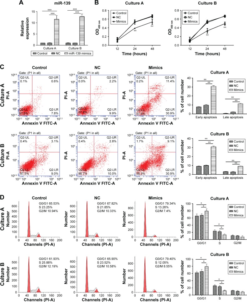
Figure 2.
miR-139-5p overexpression inhibited the proliferation of uterine leiomyoma cell and induced cell apoptosis and G1 phase arrest.
Notes: (A) miR-139-5p expression after transfection of miR-139-5p mimics was determined by RT-PCR (***P<0.001). (B) miR-139-5p overexpression inhibited the cell proliferation. Cells were treated with miR-139-5p mimics or NC. Cell growth was determined by CCK-8 assay at different time points. **P<0.01, compared with blank control and NC. (C) miR-139-5p overexpression induced cell apoptosis. Cells were treated with miR-139-5p mimics or NC and incubated for 72 hours. Apoptosis was measured by Annexin V/PI assay (**P<0.01). (D) miR-139-5p overexpression induced G1 phase arrest. Cells were transfected by miR-139-5p mimics or NC and incubated for 72 hours. Cell cycle distribution was strained by PI and determined by flow cytometry (*P<0.05).
Abbreviations: CCK-8, Cell Counting Kit-8; NC, negative control; OD, optical density; PI, propidium iodide; RT-PCR, real-time polymerase chain reaction.

