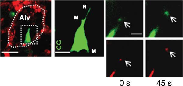
Figure 2. In vivo formation of tunneling nanotubes and microvesicles in bone-marrow derived stem cells (BM-MSC).
Mice were instilled with intranasal LPS and 4 hours after LPS, BM-MSCs were administered by intratracheal installation. We next isolated and blood-perfused the lung and imaged using confocal microscopy. (A) Image shows a mouse alveolus (red) and a mouse BM-MSC (green). The dotted oval structure represents an alveolar unit and the dotted rectangular structure demarcates a BM-MSC. Line , 10 μm. (B) Magnified image of BM-MSC as depicted in the rectangular box in figure (A). The image shows formation of nanotubes (N) and microvesicles (M). CG, calcein green. Line, 5 μm. (C) Panels show microvesicle (arrows) being released from BM-MSc at different time points in green (calcein green) and red (DsRed) renditions. Line, 3 μm. (Islam, Das et al. 2012)

