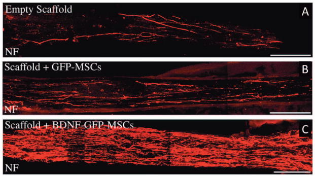
Figure 2.
Implantation of templated agarose scaffold promoted infiltration of axons into spinal cord aspiration lesion site. A) Control scaffolds promoted highly linear axonal extension through the scaffold’s longitudinal channels. B) Incorporation of brain derived neurotrophic factor secreting bone marrow stromal cells (MSC) greatly increased axonal penetration into scaffolds compared to MSC-free control scaffolds. This figure was reproduced from Stokols et al. with permission from Mary Ann Liebert Inc. [Stokols et al., 2006].

