
Figure 1.
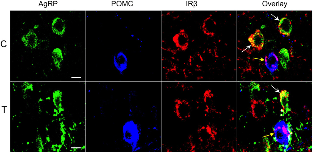
Confocal images of triple-labeled 1µm thick optical sections showing examples of AgRP- (green), POMC- (blue), and IRβ-immunolabeled neurons (red) in the arcuate nucleus of control (C, top panel) and prenatal T (T, bottom panel) ewes. Overlay images (far right panels) show colocalization of IRβ with AgRP (white arrows) and POMC (yellow arrows) neurons. Scale bars = 10µm.