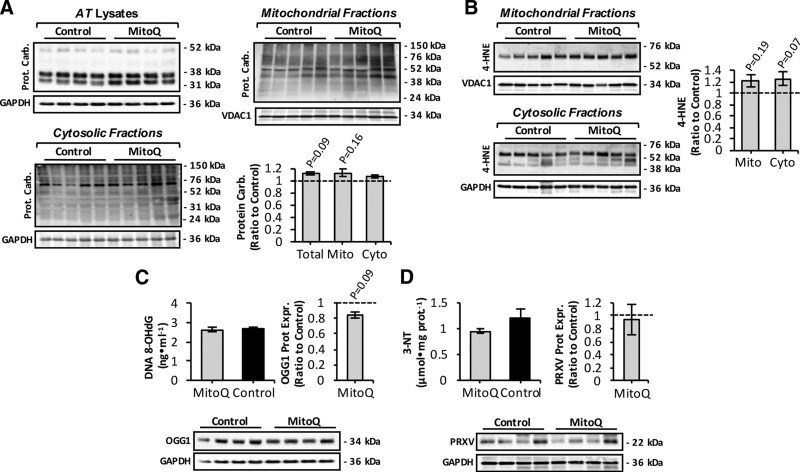
Figure 3.
Markers of oxidative damage in skeletal muscle from mitoquinone mesylate (MitoQ)-treated old mice. A) Western blot analysis and quantification (lower right) of protein carbonyls in mitochondrial (upper right) and cytosolic (lower left) skeletal muscle fractions, and AT lysates (upper left) of control and MitoQ-treated old mice. B) Western blot analysis (left) and quantification (right) of 4-HNE protein adducts in mitochondrial (upper left) and cytosolic skeletal muscle fractions (lower left) of control and MitoQ-treated old mice. C) Levels of 8-OHdG in genomic DNA extracted from skeletal muscle (upper left), and OGG1 protein levels (lower) of skeletal muscle from control and MitoQ-treated old mice and densitometric quantification of blot (n = 5–6 mice per group; upper right). D) 3-NT content (upper left) and PRXV protein levels (lower) of skeletal muscle from control and MitoQ-treated old mice and densitometric quantification of blot (n = 5–6 mice per group; upper right).

