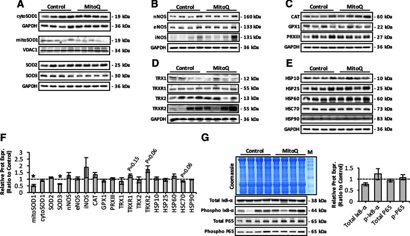
Figure 4.
Effect of long-term mitoquinone mesylate (MitoQ) treatment on RONS regulatory protein expression in skeletal muscle of old mice. A) Representative Western blots depicting SOD isoform expression in AT lysates and mitochondrial/cytosolic skeletal muscle fractions of control and MitoQ-treated old mice. B) Protein expression levels of NOS isoforms in AT lysates of control and MitoQ-treated old mice. C) Western blots of main H2O2-reducing enzymes, including catalase (CAT), glutathione peroxidase 1 (GPX1), and PRXIII, in AT lysates of control and MitoQ-treated old mice. D) Protein expression of main redox proteins involved in TRX-PRX system, including TRX1, TRX2, TRXR1, and TRXR2, in AT lysates of control and MitoQ-treated old mice. E) Western blots of heat shock proteins in AT lysates of control and MitoQ-treated old mice. F) Densitometric analysis of represented Western blots shown in A–E. *P < 0.05 compared to values from old control mice. G) Effect of long-term MitoQ treatment on total and phosphorylated IκB-α (phospho IκB-α) and P65 content (total and phosphorylated) (lower left), and densitometric quantification of blots (right). Coomassie Brilliant Blue–stained gel (upper left) served as loading control. M, molecular weight marker.

