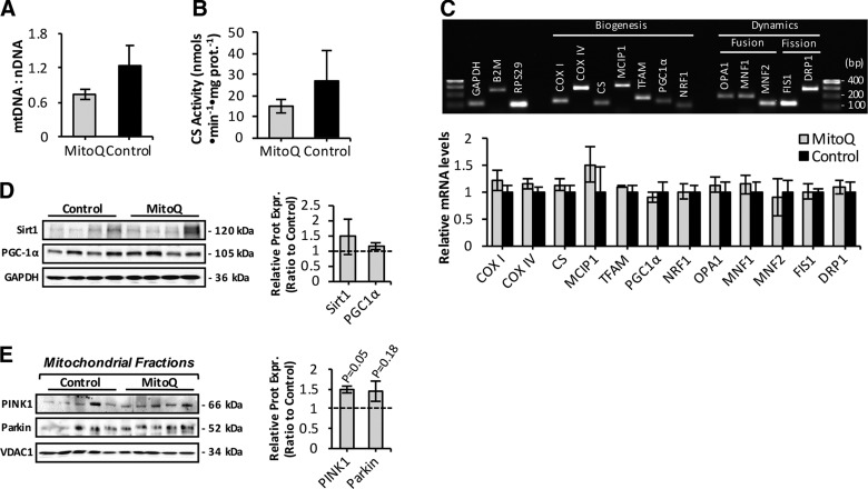
Figure 5.
Effect of long-term mitoquinone mesylate (MitoQ) administration on mitochondrial content and mitophagy in skeletal muscle of old mice. A) Real-time qRT-PCR measurement of mtDNA, normalized to amount of nuclear DNA (nDNA) in skeletal muscle of control and MitoQ-treated old mice (n = 5–6 mice per group). B) CS activity in skeletal muscle of control and MitoQ-treated old mice (n = 5–6 mice per group). C) Representative image of agarose gel electrophoresis of real-time RT-PCR amplification products of GAPDH, B2M, RPS29, COXI, COXIV, CS, MCIP1, mitochondrial transcription factor A (TFAM), PGC-1α, nuclear respiratory factor 1 (NRF1), OPA1, MNF1, MNF2, FIS1, and DRP1 transcripts (upper). Lanes 1 and 17, 100 bp DNA molecular weight marker. PCR products correspond to amplicon sizes listed in Table 1. Relative mRNA levels of genes involved in mitochondrial biogenesis and dynamics analyzed by real-time qRT-PCR (lower). mRNA levels were normalized against the housekeeping genes GAPDH, B2M, and RPS29. D) Protein expression of sirtuin 1 (Sirt1) and PGC-1α mitochondrial biogenesis regulators (left) in AT skeletal muscle of control and MitoQ-treated old mice and densitometric quantification of blots (right). E) Western blots of isolated mitochondrial fractions from skeletal muscle of control and MitoQ-treated old mice immunodetected for PINK1, and ubiquitin ligase Parkin, mitophagy markers (left), and densitometric quantification of blots (right).

