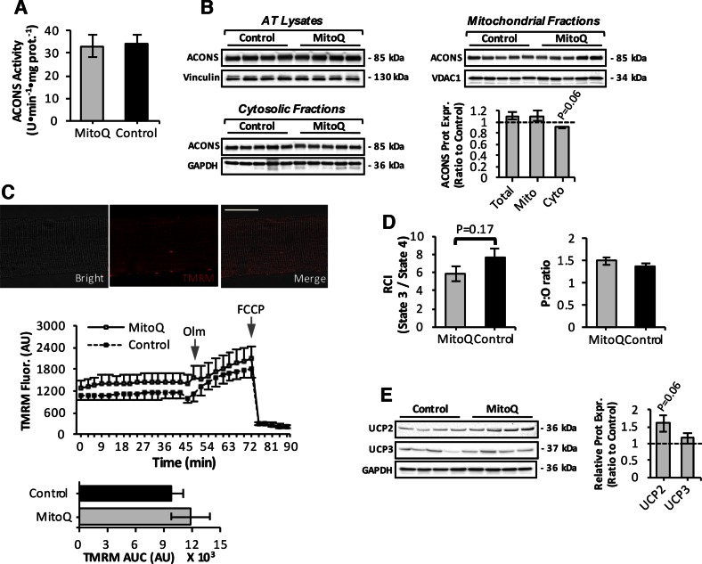
Figure 6.
Mitochondrial function in skeletal muscle of mitoquinone mesylate (MitoQ)-treated old mice. A) Mitochondrial aconitase (ACONS) activity in AT skeletal muscle of 28-mo-old control and MitoQ-treated mice (n = 5–6 mice per group). B) Western blot analysis and quantification (lower right) of ACONS in mitochondrial (upper right) and cytosolic (lower left) skeletal muscle fractions, and AT lysates (upper left) of control and MitoQ-treated old mice. C) Confocal images of single fiber isolated from AT muscle under bright-field fluorescent image after loading with TMRM fluorescence (20 nM, red); merged image as indicated and analyzed by fluorescence microscopy. Original magnification, ×60. Scale bar, 30 μm (upper); Measurement of ΔΨm in intact mitochondria of isolated AT fibers from control and MitoQ-treated old mice, assessed by changes in TMRM fluorescence in response to oligomycin (Olm; 2.5 μM) and FCCP (4 μM), added at indicated time points (center); statistical analysis of area under TMRM fluorescence trace (area under curve) for control and MitoQ-treated old mice (n = 10–12 fibers, 5–6 mice per group; lower). D) Respiratory function of intact mitochondria in saponin-permeabilized myofibers from control and MitoQ-treated old mice shown by changes in RCI (left) and ratio of ATP amount to consumed O2 during state 3 (P:O ratio) (right) (n = 5–6 mice per group). E) UCP2 and UCP3 protein levels in skeletal muscle of control and MitoQ-treated old mice (left) and densitometric quantification of blots (right).

