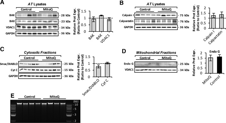
Figure 7.
Effect of long-term mitoquinone mesylate (MitoQ) treatment on mitochondrial-mediated apoptosis in skeletal muscle. A) Immunoblots of BAK, BAX, and VDAC1 proapoptotic proteins in skeletal muscle of control and MitoQ-treated old mice (left) and densitometric quantification of blots (right). B) Protein expression levels of calpain I and calpastatin proteolytic enzymes in muscle of control and MitoQ-treated old mice (left) and densitometric quantification of blots (right). C) Western blots of isolated cytosolic fractions of control and MitoQ-treated mice immunodetected for cytochrome c (Cyt C) and Smac/DIABLO mitochondrial proapoptotic proteins (left), and densitometric quantification of blots (right). D) Protein levels of proapoptotic factor endonuclease G (Endo G) in skeletal muscle mitochondrial fractions of control and MitoQ-treated old mice (left) and densitometric quantification of blot (right). E) DNA fragmentation of genomic DNA isolated from skeletal muscle of control and MitoQ-treated old mice analyzed by agarose-gel electrophoresis. Lanes 1 and 10, 1 kb DNA molecular weight marker.

