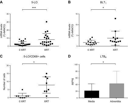
Figure 3.
Increased activation of 5-LO and LTB4–BLT1 axis in irradiated human arteries. A, B) Messenger RNA levels of 5-LO (n = 20 in each group) (A) and BLT1 (n = 12 in each group) (B) in irradiated (XRT) human arteries compared with nonirradiated (0 XRT) from the same patient. Relative gene expression results are expressed as 2–ΔCt using phosphoglycerate kinase 1 as housekeeping gene. C) Semiquantification of 5-LO and CD68+ cells was performed in 8 paired arterial biopsies after immunofluorescence staining. D) Levels of LTB4 detected in conditioned medium derived from either the adventitial or medial layer derived from irradiated arteries (n = 5 in each group). Wilcoxon’s sign-rank test of paired samples was used to test differences between XRT and 0 XRT arteries (A–C), and Student’s t test for comparisons between media and adventitia (D). Data values presented with sd. *P ≤ 0.05; ***P ≤ 0.001.

