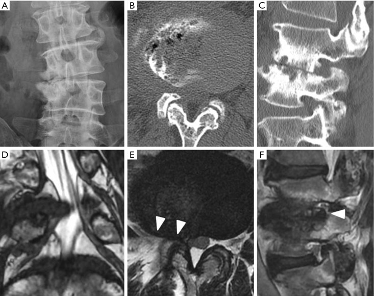
Figure 4.
Preoperative radiographic findings in a patient with degenerative scoliosis (case 4): (A) Plain anteroposterior radiograph of lumbar spine; plain CT scan: (B) axial view of L3/4 disc level, (C) sagittal view of corresponding right foramen; T2-weighted MRI: (D) coronal view showing bilateral L4 and L5 nerve roots, (E) axial view of L3/4 disc level, (F) sagittal view of corresponding right foramen. White arrowheads indicate herniated nucleus.

