. 2016 Sep 30;28(3):148–156. doi: 10.5371/hp.2016.28.3.148

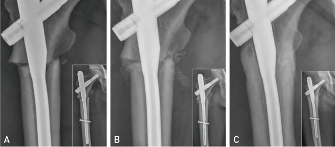
Fig. 2. The serial radiographs represent the course of atypical femoral fracture (Case 3) at subtrochanteric region with incomplete reduction. Each radiograph shows enlarged view of fracture and contains overview of proximal femur. The patient started to inject teriparatide (TPTD) after 11 days after surgery and kept using it for three months. Rapid growth of callus was observed between fracture fragments. (A) Immediate postoperative radiograph reveals slight varus reduction with a gap on lateral cortex and lack of contact on medial cortex. (B) Postoperative four-week radiograph reveals that abundant callus was formed with three weeks of TPTD injection. (C) Postoperative six month radiograph represents that the gap on both medial and lateral cortex was filled with abundant callus without trace of fracture line.

Fig. 2