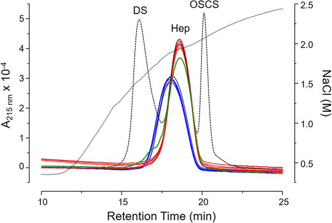
Figure 1. Anion-exchange chromatography of heparins from different animal sources.

HP-I (red line), HB-I (blue line) and HP-L (green line) were eluted from an IonPac AS11-HC column through a linear gradient of 0.4 → 2.5 M NaCl (dotted line). Dashed line shows the elution profile of a standard mixture containing 50 μg dermatan sulfate (DS), 200 μg 6th International Heparin Standard from NIBISC (Hep) and 30 μg oversulfated chondroitin sulfate (OSCS). The eluents were monitored via UV (A215nm).
