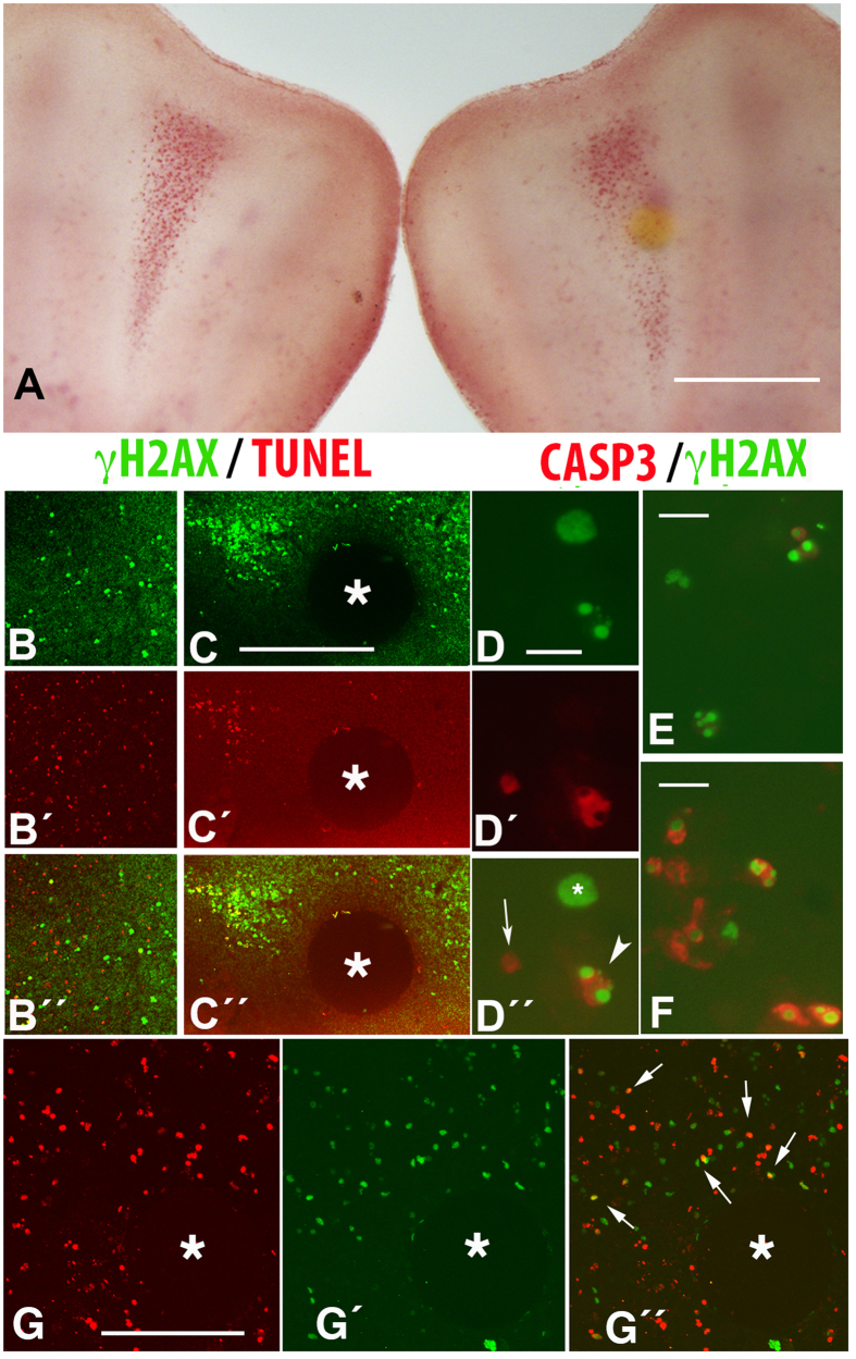
Figure 4.
(A) Control (left) and experimental (right) autopods 24 hr after the interdigital implantation of a bead incubated with the pan-caspase inhibitor and following vital staining with neutral red. Note the moderate diminution of cell death around the bead. Bar = 200 μm. (B,C) Are confocal images of control (B) and experimental (C) interdigits 24 hr after the interdigital implantation of a bead (*) bearing the pan-caspase inhibitor. Note the reduction in TUNEL-positive cells (C´; red labeling) in comparison with the contralateral, control interdigit (B´). Note also the major abundance of cells γH2AX-positive in the experimental interdigit (C, green labeling) in comparison with the control (B). B´´ and C´´ are the merged images of B–B´ and C–C´. Bar for (B,C) = 100 μm. D–D´´ Squash of the interdigital mesoderm at id 7.5 after immunolabeling with anti-γH2AX (green in D and D´´) and active caspase 3 (red in D´and D´´), to show that cells positive for γH2AX can be either negative (* in D´´) or positive (arrowhead in D´´) for caspase 3. Arrow shows a cell positive for active caspase 3 but negative for γH2AX. Bar = 15 μm. (E,F) Squashes of the third interdigit at stage 6.5 (E) and 7.5 (F) to show the increase in active caspase immunolabeling in the course of interdigit regression, Bar = 10 μm. G–G´´ are confocal pictures of the interdigital mesoderm showing TUNEL (G, red) and γH2AX (G´, green) labeling 10 hr after the implantation at id 5.5 of a H2O2 bead (*). G´´ is the merged image. Note the presence of cells positive for γH2AX only (green), cells TUNEL-positive only (red), cells double-positive for γH2AX and TUNEL (arrows in G´´). Bar = 100 μm.

