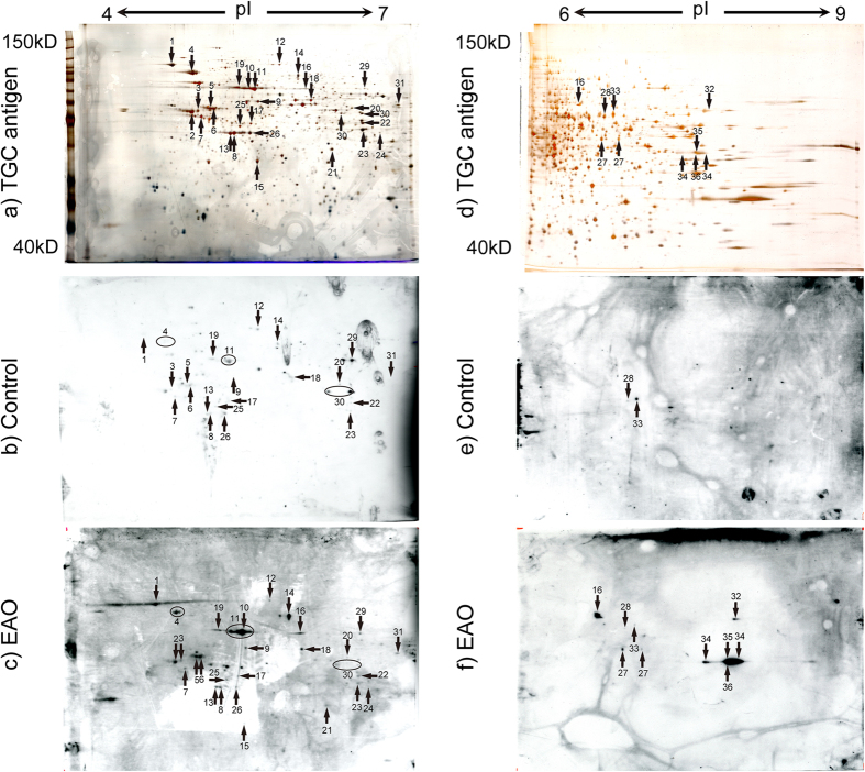
Figure 3.
Identification of AIs in TGC proteins by pI4–7 (a–c) and pI6–9 (d–f) 2D gel electrophoresis. Numbered proteins were identified on the corresponding silver-stained 2D gel with testicular proteins (a,d), immunoblotting with control serum (b,e), and immunoblotting with representative EAO serum (c,f). The numbered spots indicated proteins that were identified by MS, Tubb2c (spot no. 2), Atp6v1a (spot no. 10), Pdhb (spot no. 15), Hsc70t (spot no. 16), Fbp1 (spot no. 21), Lrrc34 (spot no. 24), Dnpep (spot no. 27), Gapdhs (spot no. 32), Pdha2 (spot no. 34), Dazap1 (spot no. 35), unnamed protein product (spot no. 36) were detected as protein relevant to EAO among the spots. The apparent molecular masses and pIs of the autoreactive proteins were determined by matching them with standard proteins with known pIs and molecular masses.

