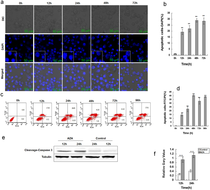
Figure 4. AZA- induced apoptosis in SL-1 cells.
(a) SL-1 cells were induced for 0 h, 12 h, 24 h, 48 h or 72 h, following staining with DAPI for 10 mins. (b) Statistical table shows the quantification of DAPI staining intensity. Data are the mean ± S.D. **p < 0.01 compared with 0 h. (c) Evaluation of apoptosis of SL-1 cells using annexin V/PI staining and flow cytometry. All these cells were treated with 2.5 μg/mL AZA for various amounts of time. Cells were double stained with annexin-V-FITC and propidium iodide (PI) and analyzed by flow cytometry. Annexin-V-, PI- cells are live cells; annexin-V+, PI- cells are early apoptotic cells and annexin-V+, PI+ cells are late apoptotic or necrotic cells. (d) Flow cytometric analysis result. Data are the mean ± S.D. **p < 0.01 compared with 0 h. (e) Western blot of cleaved-caspase-3 level induced by AZA from 12 h and 24 h in SL-1 cells. Control was without treatment. Tubulin was used as a loading control for the western blot analysis. (f) The quantification of cleaved-caspase-3 from 3 independent experiments. Data are the mean ± S.D. **p < 0.01 compared with the control group.

